Limitations of conventional mainstream approaches used to assess and treat cognitive impairment
BrainEEG.jpg

Illustration by Kateryna Kon / 123rf.com
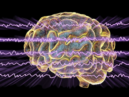
EEG Electroencephalogram, brainwave in awake state with mental activity, 3D illustration
Current pharmacologic treatments of Alzheimer’s disease (AD) work by inhibiting the enzyme that breaks down acetylcholine, thus increasing available levels of the neurotransmitter that is critical for learning and memory. Although available drugs sometimes lessen the severity of cognitive decline and behavioral disturbances that accompany AD, they do not address its root causes. Only five drugs have been approved by the U.S. Food and Drug Administration (FDA) to treat Alzheimer’s disease (AD) (Alzheimer’s Association 2019). Among these the cholinesterase inhibitors include tacrine, donepezil, galantamine and rivastigmine. Another drug, memantine, works by antagonizing glutamate receptors and is in a class by itself. Second-generation acetylcholinesterase inhibitors (donepezil, rivastigmine, and galantamine) are no more effective than tacrine but require less frequent dosing. All currently available agents have associated side effects that can be very distressing to individuals struggling with dementia, including vomiting, diarrhea and appetite loss. Tacrine, the first cholinesterase inhibitor, was removed from the market in 2013 because of concerns over hepatotoxicity.
Although neuropsychological testing and brain imaging are often helpful for evaluating memory problems and other cognitive problems, in the day-to-day practice of medicine it is often challenging to identify the causes of cognitive impairment. Available historical information is often incomplete and inaccurate, insurance coverage is limited, and the cognitively impaired individual (and his or her family) is seldom able to advocate for an appropriate and thorough evaluation. These issues are amplified by the scarcity of outpatient mental health resources available to the rapidly growing elderly population and the increasing prevalence rate of dementia in industrialized countries.
Failure to adequately characterize complex medical or psychiatric causes of cognitive impairment leads to inadequate treatment, and many patients who might otherwise benefit from treatment remain impaired. In this short piece I briefly summarize research evidence for innovative new approaches being investigated for assessment of memory loss and other problems in cognitive functioning including quantitative electroencephalography (QEEG), virtual reality tools, and measuring copper, zinc, iron and vitamin D levels in the blood.
Although currently used mainly as research tools, in the near future these assessment approaches will be used in the day to day clinical practice of mental health care, and permit mental health providers to more reliably and more accurately identify the complex causes of cognitive impairment. Advances in assessment will, in turn, allow clinicians to develop more effective treatment strategies addressing memory loss and other forms of cognitive impairment that will address the limitations of conventional pharmacologic treatments.
Emerging assessment approaches
Quantitative electroencephalography (qEEG) and neurometric brain mapping will become standard tools for predicting long-term clinical outcomes
Quantitative electroencephalography (qEEG) has been the subject of intensive research for many years. QEEG measures include power, left-right interhemispheric symmetry, and other characteristics of brain electrical activity. QEEG can be used to differentiate mild cognitive impairment (MCI) from Alzheimer’s disease (Kwak 2006). QEEG changes that take place at different stages of Alzheimer’s disease and other disorders of severe cognitive impairment reflect decreased regional brain energy metabolism or cerebral blood flow that may not be evident in other imaging studies such as computed tomography (CT) and magnetic resonance imaging (MRI) (Chiaramonti et al 1997).
Recent findings show that Alzheimer’s disease has a heterogeneous presentation and a highly variable rate of progression, and that different individuals may experience a wide range of neurologic symptoms as well as behavioral and cognitive impairments (Mangone 2004). A 2009 review of 46 studies that evaluated the diagnostic accuracy of qEEG in individuals with mild cognitive impairment (MCI) and Alzheimer’s disease found insufficient evidence of diagnostic utility of resting EEG in both conditions (Jelic & Kowalski 2009). However, more recent findings support that QEEG provides superior temporal resolution, is simpler to implement in clinical settings and more affordable than functional magnetic resonance imaging (fMRI) for analysis of brain network connectivity, and may provide a predictive neuro-marker for the risk of developing AD (Stam, 2014).
Neurometric brain mapping is a specialized qEEG approach that compares EEG characteristics of the individual being evaluated with normative databases for individuals of the same age. Neurometric mapping helps to clarify functional brain correlates of cognitive impairment, and yields information that is useful for planning EEG-biofeedback protocols addressing specific kinds of dysfunction (Surmeli et al 2016). Neurometric brain mapping is being increasingly used in clinical settings to differentiate cognitive impairments that are due to different etiologies such as head injuries, medical disorders, progressive dementia, alcohol and drug abuse, depressed mood, learning disorders, and other biological causes.
Virtual reality tools are permitting earlier, more reliable and more accurate assessment of mild cognitive impairment (MCI) and dementia
Virtual reality (VR) tools are being used to enhance the diagnostic accuracy of existing conventional neuropsychological assessment methods used to evaluate cognitive impairment in degenerative neurological disorders, stroke, developmental disorders, and traumatic brain injury (Fernandez-Montenegro & Argyriou 2017). Virtual performance testing environments are proving to be reliable and accurate for assessment of cognitive impairment compared and provide a valuable adjunct to conventional neuropsychological testing approaches (Negut 2016). Prototype VR environments have yielded promising results for assessment of memory, attention, executive functioning, sensorimotor integration, and many activities of daily living (Allain et al 2014). Innovations in Alzheimer's screening tests incorporate immersive virtual environments, concepts from interactive video games, and advanced Human Computer Interaction (HCI) systems. A VR testing environment was successfully used to differentiate individuals with mild cognitive impairment (MCI) who were more likely to progress from those who were less likely to progress at one year (Tarnanas et al 2014). VR tests have recently been shown to reliably distinguish healthy individuals from Alzheimer’s patients (Fernandez-Montenegro 2017). These tests can be used in clinical settings to assess memory loss related to common objects and recent conversations and events, evaluate deficits in expressive and receptive language, and determine an individual’s capacity to differentiate between virtual worlds and reality.
Future approaches used to assess cognitive impairment will combine VR technology with qEEG, functional MRI (fMRI) and other functional brain-imaging technologies, advancing understanding of the causes of mild cognitive impairment (MCI) and dementia. The use of VR environments to assess neuropsychological functioning will lead to individualized rehabilitation strategies that more effectively address performance deficits on a case by case basis.
Abnormal metabolism of zinc, iron and copper increase risk of developing Alzheimer’s disease
Recent findings have implicated metals such as copper, iron, and zinc as key mediating factors in the pathophysiology of Alzheimer's disease and are leading to novel treatment approaches aimed at regulating metal homeostasis in the brain (Bush & Tanzi 2008; Kenche 2011; Kumar et al 2019). The role of zinc in the pathogenesis of AD is poorly understood. Emerging findings suggest that zinc plays both a neuroprotective and disease-promoting role depending on which pathologic stressors are present (Cuajungco & Faget 2003). High levels of zinc are neurotoxic and are believed to promote formation of amyloid beta plaques, whereas histopathology studies of the brains of Alzheimer’s patients reveal deficient zinc in the brain (Cuajungco & Lees, 1997). Paradoxically, abnormal low zinc levels may also increase risk of developing AD. Low serum zinc levels result in impaired zinc transport to the brain and have been cited as a possible factor in the formation of amyloid plaques. Serologic studies of trace metals have not yet entered the domain of clinical medicine however the above findings will soon lead to routine tests of these trace metals in clinical settings, and protocols for advising individuals on supplements aimed at correcting deficiencies or excess levels of specific metals aimed at normalizing metabolic processes that contribute to Alzheimer’s disease and other degenerative neurologic disorders.
Low Vitamin D serum levels are correlated with increased risk of developing Alzheimer’s disease
Research findings support that low serum vitamin D (25-hydroxyvitamin D) levels are correlated with increased risk of developing AD. Three long-term observational studies done in different countries found that healthy elderly individuals with higher serum vitamin D levels had reduced risk of developing AD (Annweiler 2012; Afzal 2014; Littlejohns 2014). Neuroprotective effects of Vitamin D may be related to generally reduced inflammation in the body as measured by relatively lower levels of C-reactive protein (CRP) (Cannell 2014). This hypothesis is consistent with findings of elevated CRP levels in individuals with vitamin D levels below 50 nmol/L (Cannell 2014). Findings of animal studies suggest that vitamin D may prevent cell death and toxicity in aging neurons (Dursun 2011). Finally, Vitamin D may promote increased clearance of amyloid beta (Lu’o’ng 2013). It is prudent to screen all healthy adults for vitamin D levels and to recommend supplementation when levels are low. As research findings continue to accrue clinicians will start to routinely measure serum vitamin D levels and advise their patients about appropriate evidence-based vitamin D supplementation.
References
Allain, P., Foloppe, D., Beshnard, J., Yamaguchi, T., Etcharry-Bouyx, F. et al (2014) Detecting everyday action deficits in Alzheimer's disease using a nonimmersive virtual reality kitchen. J Int Neuropsychol Soc.;20(5):468-77.
Afzal S, Bojesen SE, Nordestgaard BG: Reduced 25-hydroxyvitamin D and risk of Alzheimer’s disease and vascular dementia. Alzheimers Dement 10:296–302, 2014.
Alzheimer’s Association. FDA-Approved Treatment for Alzheimer’s. at: http://www.alz.org. Accessed 7 March 2019.
Annweiler C, Herrmann FR, Fantino B, Brugg B, Beauchet O: Effectiveness of the combination of memantine plus vitamin D on cognition in patients with Alzheimer disease: a pre–post pilot study. Cogn Behav Neurol 25:121–127, 2012.
Bush, A., Tanzi, R. (2008) Therapeutics for Alzheimer's disease based on the metal hypothesis. Neurotherapeutics; 5(3):421-32.
Cannell JJ, Grant WB, Holick MF: Vitamin D and inflammation. Dermato-endocrinology 6:e983401, 2014.
Chiaramonti R, Muscas GC, Paganini M, Müller TJ, Fallgatter AJ, Versari A, Strik WK. (1997) Correlations of topographical EEG features with clinical severity in mild and moderate dementia of Alzheimer type. Neuropsychobiology. 36(3):153-8.
Chui D, Chen Z, Yu J, Zhang H, Wang W, Song Y, Yang H; Liang Zhou. (2011) Magnesium in Alzheimer’s disease. In: Vink R, Nechifor M, editors. Magnesium in the Central Nervous System, Adelaide (AU): University of Adelaide Press.
Cuajungco, M., Faget, K. (2003) Zinc takes the center stage: its paradoxical role in Alzheimer's disease. Brain Res Rev 41(1):44-56.
Dursun E, Gezen-Ak D, Yilmazer S: A novel perspective for Alzheimer’s disease: vitamin D receptor suppression by amyloid-beta and preventing the amyloid-beta induced alterations by vitamin D in cortical neurons. J Alzheimers Dis 23:207–219, 2011.
Fernandez-Montenegro, J., Argyriou, V. (2017) Cognitive evaluation for the diagnosis of Alzheimer's disease based on Turing Test and Virtual Environments. Physiol Behav. 173:42-51.
Jelic, V., Kowalski, J., (2009) Evidence-based evaluation of diagnostic accuracy of resting EEG in dementia and mild cognitive impairment. Clin EEG Neurosci. 40(2):129-42.
Kenche, V., Barnham, K., (2011) Alzheimer's disease & metals: therapeutic opportunities. Br J Pharmacol; 63(2):211-9.
Kumar, A., Sharma, P., Prasad, R., Pal, A. (2019) An urgent need to assess safe levels of inorganic copper in nutritional supplements/parenteral nutrition for subset of Alzheimer's disease patients. Neurotoxicology. 4;73:168-174.
Kwak, Y. Quantitative EEG findings in different stages of Alzheimer's disease. J Clin Neurophysiol. 2006 Oct;23(5):456-61.
Littlejohns TJ, Henley WE, Lang IA, Annweiler C, Beauchet O, Chaves PH, Fried L, Kestenbaum BR, Kuller LH, Langa KM, Lopez OL, Kos K, Soni M, Llewellyn DJ: Vitamin D and the risk of dementia and Alzheimer disease. Neurology 83:920–928, 2014
Lu’o’ng KV, Nguyen LT: The role of vitamin D in Alzheimer’s disease: possible genetic and cell signaling mechanisms. Am J Alzheimers Dis Other Demen 28:126–136, 2013.
Mangone, C. (2004) [Clinical heterogeneity of Alzheimer's disease. Different clinical profiles can predict the progression rate]. Rev Neurol. 1-15;38(7):675-81.
Negut, A., Matu, S., Sava, F., David, D. (2016) Virtual reality measures in neuropsychological assessment: a meta-analytic review. Clin Neuropsychol. 30(2):165-84.
Polich, J., Corey-Bloom, J., (2005) Alzheimer's disease and P300: review and evaluation of task and modality. Curr Alzheimer Res. 2(5):515-25.
Stam, C. J. (2014). Modern network science of neurological disorders. Nat. Rev. Neurosci. 15, 683–695.
Surmeli, T., Eralp, E., Mustafazade, I., Kos, H., Ozer, G., Surmeli, O. (2016) Quantitative EEG Neurometric Analysis-Guided Neurofeedback Treatment in Dementia: 20 Cases. How Neurometric Analysis Is Important for the Treatment of Dementia and as a Biomarker? Clin EEG Neurosci. 47(2):118-33.
Tarnanas, I., Tsolaki, M., Nef, T., et al (2014) Can a novel computerized cognitive screening test provide additional information for early detection of Alzheimer's disease? Alzheimers Dement.;10(6):790-8.


